TREATMENT OF ELBOW DYSPLASIA

Elbow Dysplasia

Elbow dysplasia refers to a group of conditions causing lameness (limping) in the front legs of dogs. Affected dogs are born with normal elbows but develop problems with the bone structure or cartilage as they grow due to abnormal bone development or uneven application of pressure to particular parts of the elbow joint. These conditions include fragmentation of the medial coronoid process of the ulna (FMCP), Ununited anconeal process (UAP), and Osteochondritis Dissecans of the humeral trochlea (OCD). Incongruity of the elbow may also be a component of the disease - this is where the bone structure of the elbow has developed abnormally and an abnormal step or gap is present between the bones. Elbow dysplasia frequently leads to osteoarthritis in affected joints and treatment is aimed at reducing the severity and progression of this. Typically affected breeds are large to giant breeds, but smaller breeds are sporadically affected.

SYMPTOMS
Symptoms of elbow dysplasia typically refer to lameness (limping) in one or both front legs. The severity of the symptoms is variable and ranges between mild, intermittent lameness and unremitting lameness that is poorly responsive to pain medication. Affected dogs frequently have effusion (swelling) of the elbow joint and pain on joint manipulation or reduced joint range of motion. Muscle bulk in the fore limb is typically reduced.

DIAGNOSIS
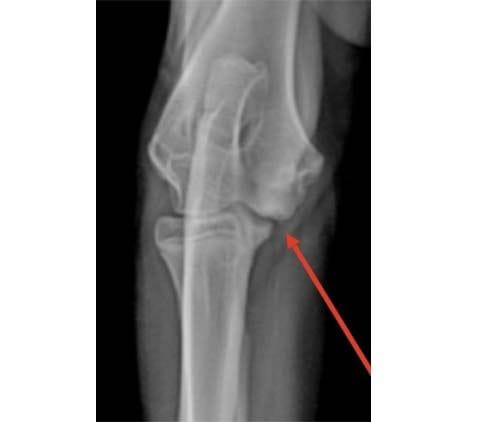
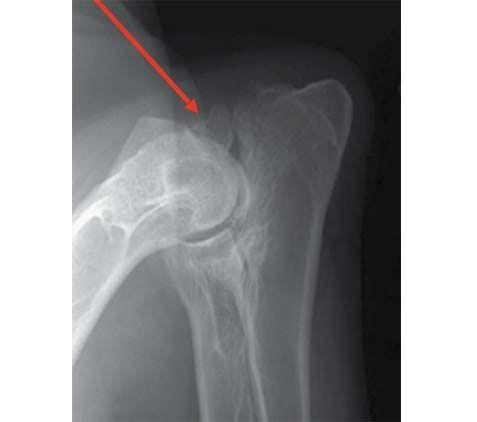
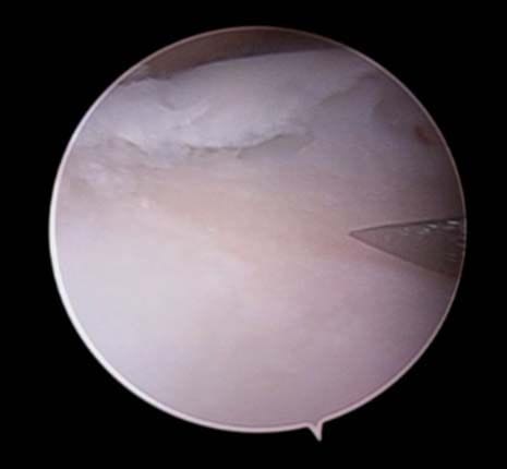
Diagnosis of elbow dysplasia is based on a combination of physical exam findings, diagnostic imaging and joint inspection via arthroscopy. Dogs affected by elbow dysplasia have pain referable to the elbow on physical examination. Radiographs can be used to diagnose UAP and OCD, as well as most types of incongruity. Fragmented medial coronoid process cannot always be definitively diagnosed by radiography due to superimposition of the radius over the medial coronoid process of the ulna, but most dogs have secondary changes to the ulna or secondary osteoarthritis that is strongly suggestive of FMCP. Computed tomography (CT) or arthroscopy (keyhole joint inspection) can be used to confirm the diagnosis. Arthroscopy allows the best assessment of the cartilage surface of the joint while CT can give information about the bone structure.

X-ray with arrow pointing to OCD lesion of distal humerus

CT image with arrow pointing at medial coronoid process fragment

X-ray with arrow pointing to
ununited anconeal process

TREATMENT
Arthroscopic (keyhole) joint inspection allows the best assessment of the cartilage surface of the joint. This is performed for most cases of elbow dysplasia. The way the condition is treated is dependent on the degree of bone fragmentation and the severity of cartilage damage. If FMCP is present, then bone fragments are removed. This fragment removal is most effective if performed in young dogs before significant arthritic change or cartilage damage occurs.

Arthroscopy picture showing medial coronoid process
fragment adjacent to arthroscopic probe

Arthroscopic picture showing exposed subchondral bone (pale pink area in fore ground with needle tip overlying) with degenerate cartilage dorsal to it.

Some cases also have microfracture (weakness) or other bone fragmentation of the remaining bone of the medial coronoid process which can be removed by arthroscopic debridement or subtotal coronoidectomy (removal of part of the medial coronoid process of the ulna). If an OCD lesion is present, the cartilage flap is removed. Severely affected dogs have cartilage loss and subchondral bone exposure despite their young age. These dogs may need further medical therapy (anti inflammatory drugs, platelet rich plasma injections, physiotherapy) or further surgery to resurface the cartilage with synthetic material (Canine Unicompartmental Elbow) or redistribute the joint load to the radius, which is usually not affected by arthritic change (PAUL procedure).

UAP is treated by screw fixation or removal of the anconeal process of the ulna, along with ulna osteotomy (bone cut in the ulna) to improve joint congruity. Some dogs have FMCP and UAP together and as such have treatment for both.

Significant incongruity is treated by ulna osteotomy in some cases, and incongruity can also coexist with other types of elbow dysplasia.

Elbow replacement systems have been developed for dogs with severe arthritis, but have not gained wide acceptance, mostly due to high complication rates and inconsistent improvement.

PROGNOSIS
Prognosis with elbow dysplasia is variable, and depends mostly on the severity of cartilage damage within the elbow joint which is assessed by arthroscopy, as well as the presence of osteoarthritis. Young dogs who are treated before the onset of secondary osteoarthritis have the best prognosis. As with any condition where damage to the joint surface occurs, osteoarthritis is a possible consequence. The severity of this is variable with some dogs managing good activity levels, while others need exercise restriction and ongoing treatment for their arthritis.

If you have any questions, please feel free to contact of the Specialist Surgeons at
Veterinary Specialist Services.

VVS
Veterinary
Hospital

Refferring A Patient?

Referring a Patient?

Find Out More
Have You been Referred?

Have You been Referred?

Find Out More