&geometry(278x56) "Veterinary Specialist Services Pty Ltd.")
Spontaneous Pneumothorax in Border Collie
 "Spontaneous Pneumothorax in Border Collie")
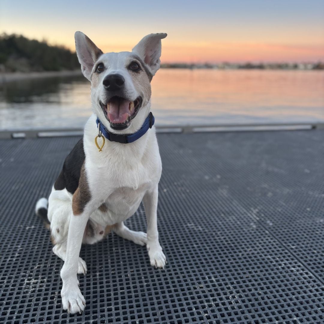
Meet Brew, an energetic, 11-year-old Border Collie Cross, who came to VSS Carrara after being referred by his regular veterinarian. His owner had noticed him being more needy over the previous two days; and on the day prior, she had noticed his breathing was short and shallow.
Brew's regular veterinarian noted these changes and recommended chest radiographs, which showed spontaneous pneumothorax - a condition where air leaks into the chest cavity, causing the lung to collapse. It's a rare, but serious condition, and in Brew's case, it was life-threatening. Causes of spontaneous pneumothorax include ruptured pneumonic bulla, abscess and tumor.
Brew was sent to the Animal Emergency Service, where he was evaluated. A chest tube was placed for removal of the air in the pleural space (the area between the chest wall and lung tissue), which allowed his lungs to better expand and stabilised his respiratory function.
He was transferred to VSS Carrara the following morning for diagnostic workup and treatment. A CT scan was recommended and this showed that Brew had a pneumonic bulla (a large air-filled blister) in the right caudal lung lobe segment. Other bullae were not seen on CT, but often they are not visible once ruptured.
Surgical exploratory was offered as the best way to treat this condition and minimize the risk of recurrence. After careful discussion with the owner, she elected to proceed. Our surgical team prepared for a median sternotomy - a procedure where the chest is opened through the breastbone to allow full access to the lungs and surrounding structures. It’s a significant operation, but one that offered the best chance of identifying and fixing Brew’s problem.
Once in surgery, we discovered the pneumonic bulla located in the right caudal lung lobe, and the lobe was removed. No additional air leaks were detected, in spite of pressure testing during the procedure. Brew recovered well, but the following day, he again developed pneumothorax. The decision was made to re-explore the chest, and a small leak was found in the right cranial lung lobe. Since there would have been significant potential for impaired respiratory ability if another lobe had to be removed, the surgeon elected to perform a surgical repair of the lung tissue. This procedure would preserve better pulmonary function; however, it is very challenging, as suturing lung tissue is very complex and can result in additional leakage in the early stages of healing.
Thankfully, Brew came through this surgery with impressive results, and in the days that followed, he recovered steadily, closely monitored by our dedicated intensive care team. It wasn't easy - chest surgery is tough, and healing takes time. However, Brew, true to his nature, faced the challenge with resilience.
Brew is now back home, surrounded by the people who love him most. He’s taking things a little slower than usual, but he’s breathing freely again and is no longer burdened by the silent danger that once threatened his life. At his first postoperative recheck two weeks later, no signs of leakage were present, he was breathing well and in high spirits. His story is a testament to the love and dedication of his owner, and her willingness to pursue the specialty care required to help him recover from such a serious ailment.
To Brew’s family: thank you for trusting us with his care. And to everyone who knows the bond between a dog and their human, Brew’s journey is a powerful reminder - our pets may grow older, but their hearts stay brave and their love never fades.
| Tags:Emergency/Critical CareMost PopularNewsPatient CareSurgery |