FRACTURE REPAIR

Fracture Fixation at Veterinary Specialist Services

In the above video, Dr Peter Delisser BVSc (Hons) Cert SAS DipECVS FHEA, European Specialist Small Animals from VSS Jindalee, talks about fracture repair options for your pet.

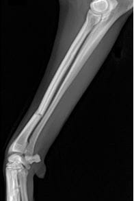
Fractures occur for a variety of reasons in dogs and cats commonly as a result of trauma or a fall. The patient may have serious concurrent internal or external injuries as a result of the causative trauma, particularly where a motor vehicle is involved. A wide variety of options are available for stabilisation of fractures depending on the type and location of the fracture.

PATIENT ASSESSMENT

Dogs and cats that are diagnosed with fractures may be investigated for internal injuries prior to surgery. This assessment may happen at the regular family veterinary practice, at an emergency clinic or with Veterinary Specialist Services. This may include radiographs (x-rays), ultrasound of the abdomen or thorax, as well as blood tests. Some animals with fractures need additional surgery or intensive medical treatment for their internal injuries prior to stabilisation of any fracture. Fractures are typically assessed with radiographs (x-rays), but in some cases a CT scan will be advised to gain more information.

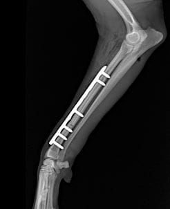
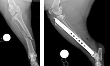
TYPES OF FRACTURE FIXATION

Veterinary Specialist Services has a wide variety of fracture fixation devices available. The exact fixation used in each fracture depends on the characteristics of the fracture, the patient age and temperament and surgeon preference. Veterinary Specialist Services has locking and dynamic compression bone plating systems, interlocking nails, circular and linear external skeletal fixation and pin/kirschner wire based fixation options along with casts and splints. In addition, fluoroscopy (low dose intra operative x-ray) is used in some procedures to confirm accurate fracture alignment and implant placement, or to minimise the extent of surgical dissection needed (minimally invasive fixation).

TIMING OF FRACTURE TREATMENT

Most fractures are best treated as soon as practically possible, providing the patient is well enough to have general anaesthesia for surgery. Dogs and cats are assessed on a case by case basis to determine the treatment plan. Initial patient management may include splinting a broken bone with a bandage or cast to support the area and provide pain relief while the patient is stabilised for surgery.

TYPES OF FRACTURES :

Long bone fractures

These are fractures of the long bones of the limbs - in the front legs the humerus, and radius and ulna, and in the back legs the femur and the tibia and fibula. These fractures are typically stabilized with bone plates, interlocking nails or external fixation, but occasionally are amenable to splint or cast support.

Pelvic Fractures

Fractures of the pelvis typically occur as a result of significant blunt force trauma such as a motor vehicle accident. These patients are more likely to have significant concurrent injuries and need additional surgery or intensive medical support prior to surgery. Pelvic fractures are treated surgically if they affect the ability of the patient to bear weight or move their legs or tail, if the patient has nerve weakness affecting the legs, tail, bladder or anal sphincter or if there are concurrent fractures to other bones in the limbs. Pelvic fractures are typically stabilized with bone plate techniques or screw fixation, and fluoroscopic assistance is frequently used.

Spinal Fractures

Spinal fractures are fractures of the bones of the vertebral column. These cases are complicated as treatment must consider both bone stabilization and nerve or spinal cord injury. Despite the severity of these injuries, most dogs recover if they retain feeling in their legs. Spinal fracture stabilisation is commonly performed with bone plates and screws, or bone screws and bone cement in combination.

Physeal or Growth Plate Fractures

Physeal fractures affect the growth plates of the bones and occur in animals before they finish growing and the skeleton matures. These growth plates are the areas of the bones that increase the length of bone as well as develop the shape of the bones at the joint surfaces. The growth plates are a relatively weak part of the bone and it therefore preferentially breaks in younger dogs compared to other mineralised parts of the bone. Growth plate fractures are typically treated as soon as possible to avoid further damage to the part of the bone responsible for growth. Growth plate fractures are often treated with pin fixation and the pins are removed in some cases to minimize the risk of subsequent growth disturbance. Growth disturbance (alteration in bone growth as a result of growth plate injury leading to altered length or angulation of a bone) can occur subsequent to physeal fracture. In most cases this is not clinically relevant, but some dogs need further surgery to correct a growth disturbance. The likelihood of growth disturbance following a growth plate fracture varies with age and type of injury, with relatively young dogs in the rapid growth phase (4-7 months depending on breed) at the highest risk.

Joint (articular) fractures

Joint fractures occur where a fracture line enters a joint. This is important as the cartilage covered joint surface has been disrupted which may have significant negative effects for joint function through poor alignment of the joint surface and subsequent development of osteoarthritis. Joint fractures are typically treated as soon as possible, and internal fixation techniques such as bone plates and screws are commonly used.

Hock and Carpal Fractures

Hock (ankle) and carpal (wrist) fractures often lead to loss of stability of the area close to the hock and carpal joints. A luxation means that a joint has dislocated - this can occur with or without a fracture. The majority of these injuries are treated with a fusion procedure called an arthrodesis. Arthrodesis involves removal of the cartilage lining of the joint and then rigid stabilisation to allow fusion of the joint space. The functional outcome of an arthrodesis depends on the joints involved. Some joints have a very small amount of movement normally and therefore stabilisation does not affect their function, while others have larger amounts of movement. Despite this large change to the amount of movement, most dogs and cats have good function following arthrodesis. A small proportion of hock and carpal fractures can be treated with screw or pin fixation of small fragments and do not require arthrodesis.

Paw (metacarpal/metatarsal) Fractures

Fractures of the paws (metacarpal or metatarsal bones and associated digits) are common and are less frequently treated with surgery. Most dogs are successfully treated with splint stabilisation for 4-8 weeks. Some animals benefit from surgical treatment.

AFTERCARE FOLLOWING TREATMENT OF FRACTURES

Patients typically stay in hospital for at least 1 night after surgical treatment, but their stay may be longer if there are severe internal injuries, or the need for longer medical support prior to discharge. Ideally, patients are comfortable on oral pain medication, and able to manage basic functions such as eating and toileting prior to discharge. For the best chance of uncomplicated bone healing, animals should be confined to a small space at home (cage, small room) until recovery is adequate and follow up x-rays show good progression of fracture healing.

PROGNOSIS FOLLOWING FRACTURE TREATMENT

Most dogs and cats return to a good level of activity following fracture repair. Each case is assessed on an individual basis and a prognosis can be discussed with the attending surgeon for each case.

If you have any questions, please feel free to contact any of the Specialist Surgeons at Veterinary Specialist Services.

VVS
Veterinary
Hospital

Refferring A Patient?

Referring a Patient?

Find Out More
Have You been Referred?

Have You been Referred?

Find Out More