&geometry(278x56) "Veterinary Specialist Services Pty Ltd.")
Cancer Across the Animal Kingdom
 "Cancer Across the Animal Kingdom")
VSS Veterinary Oncology
Beyond Dogs and Cats: Cancer Across the Animal Kingdom
A budgerigar that suddenly stops using one leg may seem an unlikely oncology patient. So might a cow with a pale patch on its eyelid or a grey horse with dark nodules beneath its tail. Yet each can be a presentation of cancer. Across the animal kingdom, the disease takes many forms, and recognising it often means looking beyond the signs familiar to dog and cat owners.
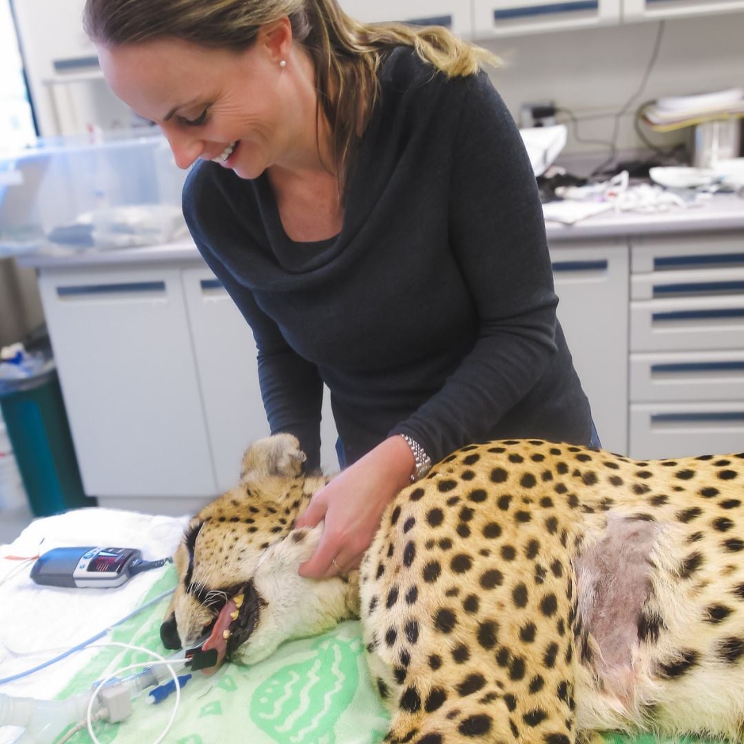
Over her career, VSS Specialist Oncologist, Dr Raelene Wouda has treated and consulted on the care of not only countless dogs and cats, but also farm animals including horses, cattle, sheep, goats, alpacas and pigs, as well as a spectrum of companion pocket pets, exotics, zoo animals and wildlife species including ferrets and prairie dogs, fish, several bird species, a giant tortoise, a bearded dragon, koalas, a wolf, a Savannah cat, a panther and two sibling cheetahs.
Each of these patients has brought the familiar questions of oncology into unfamiliar territory: What exactly is the disease? How is it likely to behave? What can reasonably be done for this animal? Across species, the answers also reveal how inherited traits, sunlight, viruses, the environment and social behaviour shape cancer.
The cases and discussions that follow draw largely on published literature, rather than Dr Wouda’s own case files. Pathology submissions, zoo records and post-mortem examinations provide valuable insights into the cancers these animals develop, while individual case reports document unusual presentations and approaches to treatment. Together, they reveal important patterns; but understanding how often cancer occurs in a species, how it behaves or how best to treat it, usually requires evidence beyond the cases available. Where species-specific evidence is limited, findings from related animals - particularly those within the same genus or family, can help inform diagnostic approaches and treatment options. These comparisons require careful clinical judgement, recognising the limitations of our knowledge base and taking into account differences in tumour behaviour, physiology and drug tolerance across species.
Cancer on Farms and in the Stable
Cancer in large animals can be readily visible on the skin or concealed within the chest, abdomen or lymphatic system. The familiar tumour names still matter, but an assessment also has to consider handling, welfare and the practical limits of treatment.
Horses: Melanoma, Sarcoids and Squamous Cell Carcinoma
Melanoma is particularly associated with ageing, grey horses. The familiar dark nodules often develop beneath the tail, around the anus/genital area or near the salivary glands behind the jaw. Around 80% of grey horses are estimated to have at least one melanoma by 15 years of age. That does not mean most will die of melanoma. Many lesions grow slowly for years, but others enlarge, interfere with defecation or tack, or spread internally. In a published series of 14 horses with metastatic melanoma, all were older grey horses; some had had skin masses for one to six years before serious internal disease was recognised. A longstanding lump can therefore deserve reassessment when it changes. Equine melanoma review; metastatic melanoma case series.
Sarcoids present a different problem. They are the most common skin tumour reported in horses and are associated with bovine papillomaviruses. They can look like a flat hairless patch, a wart or an ulcerated fleshy mass. Sarcoids are locally aggressive tumours that do not generally metastasise, but they can invade local tissue, recur after treatment and become difficult to manage beside an eye or beneath a girth. Calling them “non-metastatic” does not make them harmless. Equine sarcoid review.
Squamous cell carcinoma can affect the eye and surrounding tissues or the penis and prepuce - particularly in older horses. It may begin as a persistent sore or small raised lesion before becoming more extensive. Site and stage determine whether local treatment is possible or a larger operation is needed. These three tumour types can all begin as a visible lump, yet their causes, behaviour and treatment differ. Equine squamous cell carcinoma review.
Cattle: “Cancer Eye” and Lymphoma
Ocular squamous cell carcinoma, commonly called “cancer eye”, is a well recognised cattle cancer. It may arise on the eyelid, third eyelid or surface of the eye as a pale raised patch, then develop into a bleeding or ulcerated mass. Older cattle with unpigmented tissue around the eyes are particularly susceptible, and ultraviolet exposure contributes to risk. A small lesion may be treatable locally; a more advanced one can compromise the eye and the animal's welfare. This is a practical example of why early detection changes the choices available. Bovine ocular squamous cell carcinoma review; study of solar radiation and risk.
Lymphoma is a different and often less visible cattle cancer. Enzootic bovine leukosis is associated with bovine leukaemia virus (BLV), but most infected cattle do not develop a tumour; published reviews estimate lymphoma in fewer than 5% of infected animals. When it does occur, lymph nodes and organs including the abomasum, heart, uterus and tissues near the spinal cord may be affected. Signs depend on the site: loss of condition, digestive problems, an abnormal heartbeat or weakness can each be part of the picture. A positive BLV test is therefore not itself a lymphoma diagnosis, and a cow with lymphoma needs more than a viral test to establish what is happening. BLV epidemiology review.
Sheep, Goats and Alpacas
Small intestinal adenocarcinoma is a recognised cause of wasting in sheep in Australia and New Zealand. It grows within the bowel, so progressive loss of condition may precede any obvious mass. Another sheep cancer, ovine pulmonary adenocarcinoma, is caused by jaagsiekte sheep retrovirus in affected regions of the world. The virus transforms lung cells; the resulting tumour and fluid production can make breathing progressively difficult. The infectious agent is the virus, not cancer cells passed directly between sheep. This distinction becomes important when we consider Tasmanian devils below. Review of neoplastic wasting diseases in sheep.
Goats develop squamous cell carcinoma too, including tumours of the perineum or tail region. A retrospective study of goats seen at a veterinary teaching hospital also identified thymoma and mammary carcinoma among the more frequently diagnosed tumours. Because hospital records capture selected sick animals, they cannot tell us how often cancer occurs in the wider goat population. Goat neoplasia study; perineal squamous cell carcinoma case series.
Published reports in alpacas and llamas describe lymphoma and other malignant round cell tumours, carcinomas and sarcomas. A lymphoma involving several internal organs may initially look like a vague loss of condition. Across livestock species, investigation and treatment must account for the animal's handling, welfare, intended use and the rules governing medicines. Camelid systematic review.
Pocket Pets and Exotic Companion Animals: Small Patients, Surprising Clinical Signs
Some of these patients present with a visible mass; in others, intermittent weakness, altered behaviour or loss of appetite is the first clue. Their small size can also change how a tumour is sampled, imaged or treated.
Ferrets: A Tumour that Lowers Blood Glucose
In ferrets, a tumour may announce itself through a sudden episode of weakness, rather than a lump. Insulinoma arises from insulin-producing pancreatic cells, and episodes of low blood glucose may cause weakness, drooling, pawing at the mouth or seizures. Ferrets also develop lymphoma. Adrenal disease may be caused by hyperplasia or a tumour and often produces hair loss and hormonal signs; it should not automatically be described as malignant cancer. Review of ferret endocrinopathy.
Rabbits: More than Uterine Cancer
Uterine adenocarcinoma is a particularly important cancer in unspayed female rabbits, with risk increasing with age. In a large retrospective study of pet rabbit pathology, it was the most common tumour diagnosis, recorded in 13.1% of intact females within that study population. A rabbit may present with blood in the urine, vaginal discharge, reduced appetite or an abdominal finding, but some uterine tumours are discovered only during surgery or post-mortem examination. The frequently repeated claim that a fixed majority of all older does develop uterine cancer should not be applied uncritically across pet populations; the answer depends on which rabbits were studied and how the diagnosis was established.
Rabbits also develop lymphoma, mammary carcinoma, skin tumours and thymoma. A thymoma lies in the front of the chest and may cause breathing difficulty or prominent eyes through interference with venous drainage. The range of diagnoses is a reason to investigate a mass or unexplained decline, rather than assuming that every older rabbit with cancer has uterine disease. Spaying removes the uterus and prevents uterine adenocarcinoma, but it does not remove the need to assess other health problems. Pet rabbit pathology study; review of rabbit masses.
Rats, Mice, Hamsters, Gerbils and Guinea Pigs
In pet rats, a rapidly growing lump beneath the skin is often mammary in origin, but that does not automatically mean an aggressive carcinoma. In a study of 330 externally palpable masses submitted from pet rats, 182 arose from mammary tissue and most were benign, with fibroadenoma the most frequent diagnosis. Rat mammary tissue extends over much of the underside of the body, so a mammary tumour may appear some distance from what an owner thinks of as the chest. Rats can also develop pituitary tumours, which may present through changes in movement or behaviour rather than as a palpable mass. The submitted biopsy series tells us about lumps that were removed or sampled, not the lifetime incidence of tumours in all pet rats. Pet rat mass study; companion rat mammary tumour series.
Hamsters can develop skin and internal tumours. In a pathology series of 177 pet hamsters with tumours, 109 (62%) involved the skin or related structures, while a separate study of submitted small mammal skin masses found hair-follicle tumours, particularly trichoepitheliomas, among the hamster cases. Splenic studies in dwarf hamsters have described lymphoma and histiocytic sarcoma. These findings are a reason to examine an enlarging skin lesion or abdominal swelling, not grounds for assuming every hamster lump is malignant. Pet hamster tumour series; small mammal skin tumour study; hamster splenic lesions.
In Mongolian gerbils, changes in the ventral abdominal scent gland can produce a conspicuous midline lump. Some lesions are benign proliferations, while carcinomas have also been reported; microscopic examination is needed to distinguish them. Mice develop mammary tumours and lymphoma, but tumour patterns can vary markedly between strains and with viral exposure. Results from purpose-bred laboratory mice therefore cannot simply be presented as the expected cancer risk for a pet mouse. Gerbil scent gland study; mouse mammary tumour virus review.
Guinea pigs deserve particular attention because substantial post-mortem data are now available. A study of 2,474 pet guinea pigs examined after death identified lymphoma or leukaemia in 174 animals (7%), alongside tumours of the lungs, female reproductive tract, thyroid, skin and mammary glands. The proportion with any tumour rose markedly with age. These are findings at post-mortem, not a prediction for an individual living guinea pig. They do show that a persistent lump, progressive loss of condition or breathing difficulty may merit investigation rather than being attributed automatically to age. Guinea pig autopsy study.
Reptiles
Reptiles do develop cancer, including skin carcinomas, soft tissue sarcomas, lymphoma and leukaemia. The impression that cancer is rare in reptiles partly reflects how difficult it can be to notice illness and how few animals receive a definitive diagnosis. A retrospective study at one zoological collection identified tumours at post-mortem in 19 of 736 lizards, but those numbers reflect that institution's population and examination practices, not a universal reptile cancer rate. In a more recent teaching hospital series, skin tumours, including squamous and basal cell carcinomas, were prominent among diagnosed lizard and tortoise cases. Zoo retrospective study; teaching hospital series.
Bearded dragons provide a more detailed example. A series of five dragons with lymphoid leukaemia described vague signs such as lethargy, reduced appetite, weight loss and ocular discharge. Blood tests, rather than a visible mass, helped reveal the disease. More recent reports used tissue markers such as CD3 to establish that some of these cancers arose from T lymphocytes. In one case, cancerous cells infiltrated multiple organs, including the liver, spleen, lungs and tissues around the nervous system. These reports demonstrate that “lymphoma” or “leukaemia” is only the beginning of a diagnosis: lineage, distribution and behaviour matter, yet treatment data in dragons remain very limited. Five-dragon series; immunophenotyping report; systemic T-cell case.
A published radiated tortoise case illustrates a different challenge. A large forelimb soft tissue sarcoma restricted movement and prevented the tortoise from withdrawing that limb. Surgeons amputated it and attached a custom support to the shell to aid mobility. The result is striking, but it is one case, not evidence that amputation will be suitable for every tortoise. An animal's ability to move, feed and behave normally is integral to judging whether a treatment has succeeded. Tortoise case report.
Beneath the Feathers
Birds belong together here because the same diagnostic problem runs from a pet budgerigar to a zoo penguin: a tumour may first appear as a change in movement, feeding or feather care, rather than as an obvious lump. An Australian clinic study examined tissue from 141 companion and aviary birds with suspected solid tumours over three and a half years. It found that cytology could be a useful preliminary test, although a tissue diagnosis remains important when the tumour type will change treatment. This was a selected clinical population, not a measure of cancer risk across all birds. Melbourne avian tumour survey.
Budgerigars: When a Limp Begins at the Kidney
A budgerigar with a kidney tumour may be brought to the vet because it is lame. The kidneys lie close to nerves supplying the legs, and a mass can affect one leg before an owner suspects an internal problem. In a study of 74 budgerigars investigated for suspected abdominal tumours, 47 had kidney tumours; reproductive tract and liver tumours were also identified, sometimes together. The striking proportion reflects this selected group of birds with suspected abdominal disease, not the risk for every pet budgerigar. Budgerigar tumour study; renal neoplasia investigation.
Parrots: A Cancer that Interferes with Eating
Squamous cell carcinoma can arise within a bird's mouth or digestive tract, where an early lesion may be mistaken for inflammation or infection. In a series of 12 parrots with alimentary squamous cell carcinoma, the oral cavity was the most frequent site, affected in six. The tumours varied in appearance and the reported signs were often nonspecific. A bird that drops food, has difficulty swallowing or loses weight may need an oral examination and sampling rather than repeated treatment for a presumed infection. Psittacine squamous cell carcinoma series.
Penguins: A Chronic Gland Problem with a Serious Differential
Penguins have a preen gland near the tail that helps maintain their plumage. A zoo case series described squamous cell carcinoma of this gland in five chinstrap and gentoo penguins. Most were older than ten years and had a history of recurrent gland problems, including impaction, rupture or abscesses. Chronic inflammation was proposed as a possible contributor, not proved as the cause. The clinical lesson is more immediate: a repeatedly affected gland warrants reassessment and, where appropriate, biopsy, because a familiar-looking lesion can conceal a different diagnosis. Penguin preen-gland case series.
Zoo Animals and Conservation
Zoo populations offer opportunities for careful observation and post-mortem study, often over an animal’s entire life. Yet a cancer diagnosis still belongs to an individual whose comfort and behaviour matter. For a managed population, it may also influence reproductive planning, particularly if disease appears before an animal has bred. Big cats, prairie dogs and black-footed ferrets illustrate different parts of that balancing act.
Big Cats
Two published cheetah cases show why the anatomical extent and the identity of a cancer both matter. One animal had T-cell lymphoma apparently confined to the liver, diagnosed with tissue markers that distinguished it from other round cell tumours. Another had multicentric T-cell lymphoma and a separate cutaneous haemangiosarcoma: two cancers in the same patient, each requiring its own diagnosis. Case reports establish that these presentations occur, but offer little basis for predicting the next cheetah’s response to treatment. Hepatic lymphoma report; multicentric lymphoma report.
Broader zoo records add context. One necropsy study of 38 captive wild felids found tumours in 19, with tumour findings increasing with age; that proportion belongs to this selected group of animals examined after death. A larger archive and literature review catalogued 554 neoplasms across 20 nondomestic felid species in one pathology archive, plus 984 published neoplasms in the same species. It identified recurring diagnoses across big cats, including mammary carcinoma and lymphoma, while showing how uneven the evidence remains between species. Wild felid necropsy study; nondomestic felid tumour review.
Mammary carcinoma also raises a management question beyond the immediate patient. A pathology study compared mammary cancers in zoo felids with and without exposure to the progestin contraceptive melengestrol acetate (MGA). Exposure was associated with tumour occurrence in that study, while the cancers looked and behaved similarly in treated and untreated animals. Such evidence informs how a breeding programme weighs contraception against possible long-term effects; it does not identify the cause of an individual animal’s cancer. Zoo felid mammary carcinoma study.
Black-tailed Prairie Dogs
A retrospective tumour series examined black-tailed prairie dogs. In 50 animals whose tumours were submitted to a zoological pathology service, the more frequent diagnoses included liver adenomas and carcinomas and lymphoid malignancies. Elodontoma, a tumour-like dental growth, was also recorded, illustrating why a pathology series of “tumours” does not consist solely of cancers. The liver, gut, blood and lymphatic tissues, and skin were among the affected systems. A separate report described anaplastic large T-cell lymphoma in three black-tailed prairie dogs. Prairie dog tumour series; T-cell lymphoma report.
Black-footed Ferrets
The black-footed ferret is an endangered mustelid. In a study of adult animals in a captive breeding programme, apocrine gland, kidney tubular cell and biliary tumours featured prominently. The likelihood of several tumour types rose with age. The authors observed that many clinically important tumours occurred after the breeding age, a useful reminder that cancer frequency in long-lived captive animals and its effect on a wild population are not necessarily the same question. Black-footed ferret study; renal tumour series.
Australian Native Wildlife
The questions widen again in Australian wildlife. A tumour may prevent one animal from feeding or breeding, while an infectious process can affect many animals over time. Koalas and sea turtles show how viruses may be associated with tumour development. Tasmanian devils reveal something rarer: a cancer whose living cells pass between animals.
Koalas and Koala Retrovirus
Koalas can develop lymphoma and leukaemia, and koala retrovirus (KoRV) is associated with increased cancer risk. Unlike most viruses discussed in everyday veterinary practice, KoRV is in the process of becoming incorporated into the inherited genome of some koala populations. Researchers studying tumour and healthy tissues from ten koalas mapped viral integration sites and found clusters near genes involved in cancer. That finding offers a plausible mechanism by which integration could contribute to tumour development; it does not mean that every infected koala will develop cancer or that all koala lymphomas have the same cause. It also makes koalas an unusually informative species for studying a process that happened much further in the past in the ancestry of many mammals. Koala retrovirus and cancer study.
Green Sea Turtles
Green sea turtles develop fibropapillomatosis, which can produce multiple growths on their skin, eyes, flippers and around the mouth. A growth in the wrong place can impair vision, swimming or feeding, regardless of its microscopic classification. Chelonid alphaherpesvirus 5 is closely associated with the condition and has been studied at Queensland foraging grounds. Researchers have also examined why tumour prevalence differs along the Queensland coast and whether water quality contributes. Viral infection alone does not explain which turtles develop visible disease, how severe it becomes or why some locations are more affected than others. For a rehabilitating turtle, the immediate question may be whether growths can be removed and the animal can feed and swim well enough to return to the sea. For conservation researchers, the same diagnosis raises questions about habitat and population health. Queensland turtle virus study; Queensland prevalence and water quality study.
Tasmanian Devils: When the Cancer Itself Spreads
Tasmanian devil facial tumour disease is different from a virus-associated cancer. Devils bite one another during feeding and mating interactions. If living tumour cells from an affected devil enter a wound in another, those cells may survive and grow as a new tumour. The new growth is descended from the original animal's cancer, rather than arising independently from the bitten devil's own cells. Tumours around the mouth and face can make eating difficult, with consequences for survival and reproduction. Original allograft report.
Ordinarily, an immune system should recognise cells from another animal as foreign. Research on the first devil tumour lineage, DFT1, found that its cells reduce expression of surface molecules important for immune recognition, helping them evade that defence. Genetic work then uncovered something even more surprising: a second, independently arisen transmissible cancer, DFT2. These are two separate cancer cell lineages spreading through the same species, not one tumour that simply changed its name. Immune evasion study; DFT2 discovery; genomic comparison.
The disease has caused severe declines in some devil populations. Work on immune recognition has informed experimental vaccination and immunotherapy research, while conservation programmes also manage populations and movement of animals. These are developing strategies, not an established cure for a wild devil with facial tumour disease. The case illustrates a challenge that individual clinical oncology cannot solve alone: a cancer that persists and evolves as it moves through a population. Vaccine research review.
Life in the water
Cancer in an individual pet fish calls for a diagnosis and a decision about its care. In wild fish and marine mammals, patterns of disease may also raise questions about an entire environment. Those scales of enquiry are connected, but they require different kinds of evidence.
From a Koi’s Skin Tumour to Cancer in Wild Fish
A lump on a fish can be a true tumour, but its appearance alone cannot distinguish cancer from infection, inflammation or a cyst. A recent report of a koi with a skin mass used examination of collected cells, tissue architecture and even electron microscopy to diagnose a cutaneous fibrosarcoma. It shows that the familiar tools of oncology can apply underwater, although anaesthesia, sampling, surgery and recovery must be adapted to fish physiology and water quality. Koi fibrosarcoma report.
Fish also reveal something larger than the individual patient. In Puget Sound in the United States, studies of bottom-dwelling English sole found liver tumours and earlier liver changes more often at contaminated sites. Researchers linked these patterns to aromatic hydrocarbons in sediments through several lines of evidence, including chemical exposure measures and experimental work. The findings make fish useful sentinels of environmental damage. They do not mean that every tumour in a pet fish was caused by polluted water, or that an association at one site can be applied unchanged to another species and place. English sole liver tumour investigation; multi-species contaminant study.
The distinction between benign and malignant disease is also important here. Fish develop many kinds of growths in skin and internal organs, and some tumour-like lesions are associated with infectious agents. Establishing a diagnosis may require cytology, biopsy or post-mortem examination. Even when a tumour can be named, evidence for treatment and prognosis in a particular fish species may consist of a handful of case reports rather than a clinical trial. Review of neoplasia in fishes.
Cancer in free-ranging marine mammals is especially difficult to study. Even finding an affected animal can be unusual, and its exposure history is seldom known. Sustained study of particular populations has nevertheless raised important questions.
Sea Lions and Beluga Whales
California sea lions are affected by urogenital carcinoma, a cancer of the reproductive tract that may invade locally and spread. In one study of sea lions examined after stranding and death at a rehabilitation centre between 2005 and 2015, 14% had cancer and most of those cancers were urogenital carcinoma. This is a striking finding in a selected group of stranded animals, not the prevalence in all wild sea lions. Otarine herpesvirus 1 is strongly associated with these tumours, while studies have also investigated genetic susceptibility and persistent environmental contaminants. The virus has been found in some sea lions without cancer, so association should not be presented as proof that it acts alone. Genetic analysis has not supported direct transfer of the cancer cells between sea lions, as occurs in Tasmanian devils. Stranded sea lion pathology study; sea lion cancer review; virus study; test of tumour-cell transmission.
Researchers have also documented an unusual burden of cancer, including intestinal cancers, in beluga whales from Canada's St Lawrence Estuary. Industrial contaminants have been investigated as a possible contributor, and later work has examined DNA damage associated with polycyclic aromatic hydrocarbons. This concerns a particular, intensively studied population. It does not establish that all beluga populations have the same cancer risk, nor that one pollutant has been proved to cause every case. St Lawrence beluga cancer study; DNA damage study.
What Other Species Can Teach Us About Cancer
The cases above raise a question that reaches beyond a catalogue of tumour types: why does the same basic process, cells escaping the controls on growth and survival, unfold so differently across species? Why do grey horses so often develop melanomas that may remain indolent for years? Why can one virus raise cancer risk in koalas while another creates a contagious lung cancer in sheep? How does a Tasmanian devil tumour survive as a living cell lineage after passing into a new host? Answers to these questions may reveal biological mechanisms that would be difficult to see by studying one species alone.
Cancer resistance can be equally informative. Naked mole-rats live remarkably long lives for their size, yet cancer appears uncommon. Laboratory research has linked part of that resistance to exceptionally large hyaluronan molecules in the material surrounding their cells. Early descriptions sometimes called them cancer-free, but several spontaneous cancers were reported in 2016. Their biology is striking precisely because the resistance is strong rather than absolute. Hyaluronan research; first reported cancers; additional cases.
Elephants pose another puzzle. An animal with so many cells and such a long lifespan might be expected to face a substantial cancer burden, yet comparative studies have found protective mechanisms, including multiple copies of the tumour-suppressor gene TP53 and a distinctive cellular response to DNA damage. At a very different scale, soft-shell clams can develop a leukaemia in which cancer cells pass through seawater to another animal. Neither discovery is a ready-made treatment for pets or people. Both broaden the questions researchers can ask about how tumours arise, evade defences and persist. Cancer risk across mammals; elephant study; clam study.
What we still lack is often as revealing as what we know. A single tortoise case can show that surgery is possible, but says little about recurrence or whether another tortoise would benefit. Differences between species may reflect biology, lifespan, environmental exposure or simply who receives veterinary care and a post-mortem examination. To turn remarkable observations into dependable advice, we need species-specific records, accurate pathology, molecular studies and treatment outcomes that include the animal’s quality of life.
That work can benefit both veterinary and human medicine. Methods developed for human cancer have helped veterinary pathologists classify tumours and investigate their mechanisms. Naturally occurring cancers in animals can, in turn, expose the roles of viruses, immune recognition, inherited defences and shared environments in ways that laboratory models may miss. A similarity between an animal tumour and a human cancer is a starting point for research, not a promise of a treatment; discoveries must be tested carefully in the species that would receive it.
For the horse with a changing melanoma, the budgerigar that can no longer use a leg or the koala with lymphoma, the first obligation is still to understand and care for that individual. Following those cases with curiosity and rigour may also answer larger questions: why some cancers become common, why others scarcely arise, and which defences might one day help animals and people alike.
References
1. Equine melanoma updates. https://pubmed.ncbi.nlm.nih.gov/39266414/
2. Metastatic melanoma in horses. https://pubmed.ncbi.nlm.nih.gov/12141308/
3. Association of bovine papillomavirus with the equine sarcoid. https://pubmed.ncbi.nlm.nih.gov/12692268/
4. Squamous cell carcinomas in horses: an update of the aetiopathogenesis and treatment options. https://pubmed.ncbi.nlm.nih.gov/39168748/
5. Bovine ocular squamous cell carcinoma. https://pubmed.ncbi.nlm.nih.gov/21056799/
6. Association between solar radiation and ocular squamous cell carcinoma in cattle. https://pubmed.ncbi.nlm.nih.gov/1854107/
7. Current developments in the epidemiology and control of enzootic bovine leukosis as caused by bovine leukemia virus. https://pubmed.ncbi.nlm.nih.gov/33352855/
8. Neoplasia-associated wasting diseases with economic relevance in the sheep industry. https://pubmed.ncbi.nlm.nih.gov/33546178/
9. Goats diagnosed with neoplasia display variability in clinical presentations, treatments, and outcomes. https://pubmed.ncbi.nlm.nih.gov/36913386/
10. Presentation, treatment, and outcome of squamous cell carcinoma in the perineal region of 9 goats. https://pubmed.ncbi.nlm.nih.gov/26483578/
11. Neoplasia and proliferative lesions of New World camelids: a systematic literature review and retrospective study of cases submitted to Colorado State University from 1995 to 2020. https://pmc.ncbi.nlm.nih.gov/articles/PMC8569619/
12. Endocrinopathy and aging in ferrets. https://doi.org/10.1177/0300985815623621
13. Neoplasia and tumor-like lesions in pet rabbits (Oryctolagus cuniculus): a retrospective analysis of cases between 1995 and 2019. https://pubmed.ncbi.nlm.nih.gov/33213301/
14. Not just uterine adenocarcinoma: neoplastic and non-neoplastic masses in domestic pet rabbits (Oryctolagus cuniculus): a review. https://journals.sagepub.com/doi/10.1177/03009858211002190
15. What ails the pet rat? A retrospective study of more than 300 biopsy samples of externally palpable masses submitted from pet rats for histopathology (2013-2021). https://pubmed.ncbi.nlm.nih.gov/38772057/
16. Description of the prevalence, histologic characteristics, concomitant abnormalities, and outcomes of mammary gland tumors in companion rats (Rattus norvegicus): 100 cases (1990-2015). https://pubmed.ncbi.nlm.nih.gov/27823365/
17. Tumours in 177 pet hamsters. https://pubmed.ncbi.nlm.nih.gov/33646624/
18. Cutaneous and subcutaneous tumours of small pet mammals: retrospective study of 256 cases (2014-2021). https://pubmed.ncbi.nlm.nih.gov/35454212/
19. Retrospective pathological studies of splenic lesions in domestic hamsters (Phodopus spp.). https://pubmed.ncbi.nlm.nih.gov/30360911/
20. Surgical excision and morphological evaluation of altered abdominal scent glands in Mongolian gerbils (Meriones unguiculatus). https://doi.org/10.1136/vr.100380
21. Mouse mammary tumor virus (MMTV) and MMTV-like viruses: an in-depth look at a controversial issue. https://pubmed.ncbi.nlm.nih.gov/35632719/
22. Neoplasia in pet guinea pigs: a retrospective analysis of 2,474 autopsy examinations. https://pmc.ncbi.nlm.nih.gov/articles/PMC11559762/
23. Reptile neoplasia at the Philadelphia Zoological Garden, 1901-2002. https://pubmed.ncbi.nlm.nih.gov/17312806/
24. Prevalence, diagnosis, treatment outcomes and immunohistochemical characterization of neoplastic disorders in reptiles presented at a veterinary teaching hospital: a cross-sectional study (2010-2023). https://pubmed.ncbi.nlm.nih.gov/40306459/
25. Lymphoid leukemia in five bearded dragons (Pogona vitticeps). https://pubmed.ncbi.nlm.nih.gov/33754817/
26. Pax5 and CD3 immunophenotyping of lymphoma in 2 central bearded dragons. https://pubmed.ncbi.nlm.nih.gov/35264043/
27. Systemic CD3+ T-cell lymphoblastic leukemia in a bearded dragon (Pogona vitticeps): clinical, therapeutic, and pathological findings. https://doi.org/10.3390/ani15182736
28. Undifferentiated sarcoma resolved by forelimb amputation and prosthesis in a radiated tortoise (Geochelone radiata). https://pubmed.ncbi.nlm.nih.gov/17315468/
29. Survey of the clinical, cytological, and histopathological features associated with neoplasms in captive avian species in Melbourne, Australia. https://pubmed.ncbi.nlm.nih.gov/37962318/
30. Neoplasms in budgerigars (Melopsittacus undulatus): clinical, pathomorphological and serological findings with special consideration of kidney tumours. https://pubmed.ncbi.nlm.nih.gov/18766792/
31. Investigations on the diagnosis and retroviral aetiology of renal neoplasia in budgerigars (Melopsittacus undulatus). https://pubmed.ncbi.nlm.nih.gov/20544420/
32. Alimentary squamous cell carcinoma in psittacines: 12 cases and review of the literature. https://pubmed.ncbi.nlm.nih.gov/34078200/
33. Uropygial gland squamous cell carcinoma in chinstrap (Pygoscelis antarcticus) and gentoo (Pygoscelis papua) penguins at the Wildlife Conservation Society's Central Park Zoo. https://pubmed.ncbi.nlm.nih.gov/25831583/
34. Solitary T-cell hepatic lymphoma with large granular lymphocyte morphology in a captive cheetah (Acinonyx jubatus). https://pubmed.ncbi.nlm.nih.gov/26056904/
35. Multicentric T-cell lymphoma and cutaneous hemangiosarcoma in a captive cheetah (Acinonyx jubatus). https://pubmed.ncbi.nlm.nih.gov/26667562/
36. Pathology in captive wild felids at German zoological gardens. https://pubmed.ncbi.nlm.nih.gov/26086731/
37. A retrospective study of neoplasia in nondomestic felids in human care, with a comparative literature review. https://pubmed.ncbi.nlm.nih.gov/34130383/
38. Histologic features of mammary carcinomas in zoo felids treated with melengestrol acetate (MGA) contraceptives. https://pubmed.ncbi.nlm.nih.gov/17491073/
39. A retrospective study of tumours in black-tailed prairie dogs (Cynomys ludovicianus) submitted to a zoological pathology service. https://www.sciencedirect.com/science/article/abs/pii/S0021997512000199
40. Anaplastic large T-cell lymphoma in three black-tailed prairie dogs (Cynomys ludovicianus). https://pmc.ncbi.nlm.nih.gov/articles/PMC5487801/
41. Epidemiology of neoplasia in captive black-footed ferrets (Mustela nigripes), 1986-1996. https://pubmed.ncbi.nlm.nih.gov/12462486/
42. Renal tubular-cell neoplasms in black-footed ferrets (Mustela nigripes): 38 cases. https://pubmed.ncbi.nlm.nih.gov/16672574/
43. Retroviral integrations contribute to elevated host cancer rates during germline invasion. https://www.nature.com/articles/s41467-021-21612-7
44. Molecular evidence for horizontal transmission of chelonid alphaherpesvirus 5 at green turtle (Chelonia mydas) foraging grounds in Queensland, Australia. https://journals.plos.org/plosone/article?id=10.1371/journal.pone.0227268
45. Spatial distribution of fibropapillomatosis in green turtles along the Queensland coast and an investigation into the influence of water quality on prevalence. https://doi.org/10.1111/csp2.12755
46. Allograft theory: transmission of devil facial-tumour disease. https://pubmed.ncbi.nlm.nih.gov/16452970/
47. Reversible epigenetic down-regulation of MHC molecules by devil facial tumour disease illustrates immune escape by a contagious cancer. https://pubmed.ncbi.nlm.nih.gov/23479617/
48. A second transmissible cancer in Tasmanian devils. https://pubmed.ncbi.nlm.nih.gov/26711993/
49. The evolution of two transmissible cancers in Tasmanian devils. https://pmc.ncbi.nlm.nih.gov/articles/PMC7614631/
50. Devil facial tumours: towards a vaccine. https://www.tandfonline.com/doi/abs/10.1080/08820139.2019.1624770
51. Cytologic, histologic, and ultrastructural findings of spontaneous, cutaneous fibrosarcoma in a koi. https://doi.org/10.1111/vcp.70096
52. Overview of studies on liver carcinogenesis in English sole from Puget Sound; evidence for a xenobiotic chemical etiology. I: Pathology and epizootiology. https://pubmed.ncbi.nlm.nih.gov/2360038/
53. Relationships between toxicopathic hepatic lesions and exposure to chemical contaminants in English sole (Pleuronectes vetulus), starry flounder (Platichthys stellatus), and white croaker (Genyonemus lineatus) from selected marine sites on the Pacific Coast, USA. https://pubmed.ncbi.nlm.nih.gov/8033852/
54. Neoplasia in fishes. https://pubmed.ncbi.nlm.nih.gov/15296871/
55. Prevalence of urogenital carcinoma in stranded California sea lions (Zalophus californianus) from 2005-15. https://pubmed.ncbi.nlm.nih.gov/29498901/
56. Common cancer in a wild animal: the California sea lion (Zalophus californianus) as an emerging model for carcinogenesis. https://pubmed.ncbi.nlm.nih.gov/26056370/
57. Unlocking the role of a genital herpesvirus, otarine herpesvirus 1, in California sea lion cervical cancer. https://pubmed.ncbi.nlm.nih.gov/33668446/
58. No evidence for clonal transmission of urogenital carcinoma in California sea lions (Zalophus californianus). https://pubmed.ncbi.nlm.nih.gov/28948233/
59. Cancer in wildlife, a case study: beluga from the St. Lawrence estuary, Québec, Canada. https://pubmed.ncbi.nlm.nih.gov/11882480/
60. Intestinal polycyclic aromatic hydrocarbon-DNA adducts in a population of beluga whales with high levels of gastrointestinal cancers. https://pmc.ncbi.nlm.nih.gov/articles/PMC6320268/
61. High-molecular-mass hyaluronan mediates the cancer resistance of the naked mole rat. https://www.nature.com/articles/nature12234
62. Initial case reports of cancer in naked mole-rats (Heterocephalus glaber). https://pubmed.ncbi.nlm.nih.gov/26846576/
63. Four cases of spontaneous neoplasia in the naked mole-rat (Heterocephalus glaber), a putative cancer-resistant species. https://pubmed.ncbi.nlm.nih.gov/27129918/
64. Cancer risk across mammals. https://www.nature.com/articles/s41586-021-04224-5
65. Potential mechanisms for cancer resistance in elephants and comparative cellular response to DNA damage in humans. https://pubmed.ncbi.nlm.nih.gov/26447779/
66. Horizontal transmission of clonal cancer cells causes leukemia in soft-shell clams. https://pubmed.ncbi.nlm.nih.gov/25860608/
| Tags:Diagnostic TestingEndocrinologyInternal MedicineMost PopularNewsOncologyPathologyPatient Care |